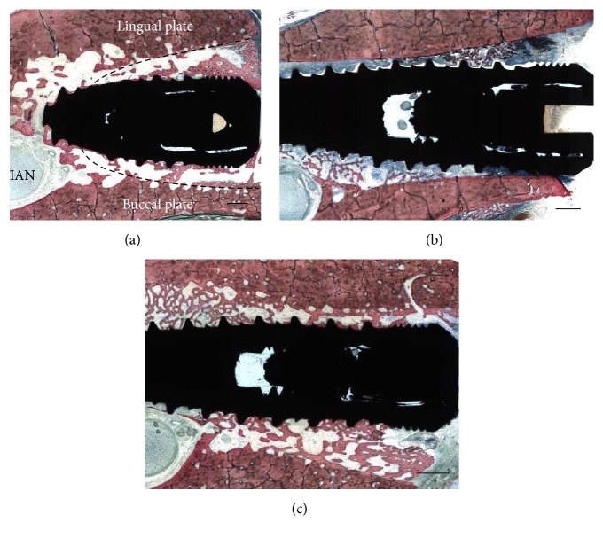
Figure 2.
(a) Observation of histologic sections depicted the implant placed in the center of the socket (estimated by the dotted line) in proximity with the inferior alveolar nerve (IAN). The gap distance at the time of implant placement between the implant and buccal/lingual plates was also observed at six weeks and presented newly formed bone partially filling this gap. Bar = 2 mm. Representative histologic sections for implants placed in sockets (b) without L-PRF and (c) with L-PRF. The absence of PRF around the implant in (a) resulted in partial soft tissue apical migration in the gap comprised by the implant and extraction socket wall, while soft tissue apical migration in (b) was avoided by the presence of the L-PRF scaffold. No notable differences in socket healing pattern were observed between surfaces regardless of implant placement with or without L-PRF. Bar = 3 mm for (b and c).

