
Figure 7.
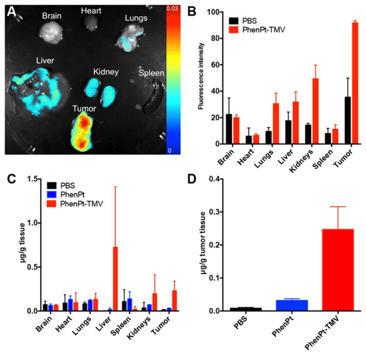
Biodistribution of PhenPt-TMV in MDA-MB-231 tumor bearing animals. (A) Representative Maestro imaging of excised organs 24 h after administration Cy5-labeled PhenPt-TMV. (B) Quantitative ROI analysis of excised organs from (A) quantifying average fluorescence intensity (tissues from n = 3 animals were analyzed). (C,D) Platinum concentration in organ tissue as measured by atomic absorption spectroscopy 24 h post-administration of phenanthriplatin or PhenPt-TMV followed by tissue homogenization.