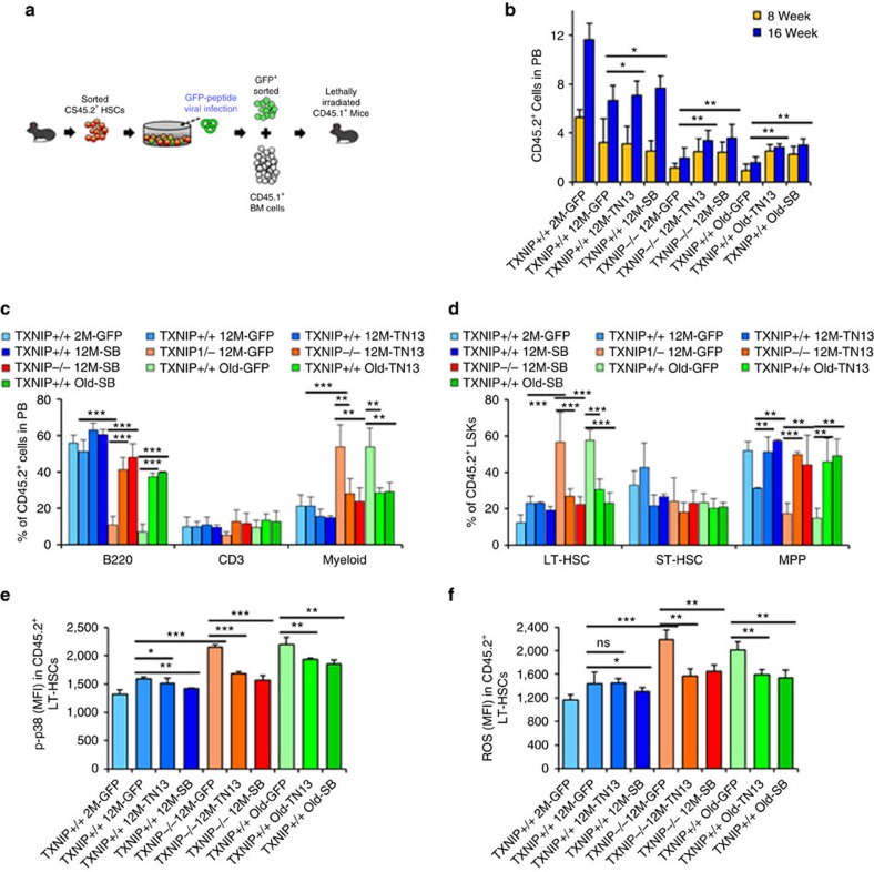
Figure 5. Rejuvenation of aged HSCs by GFP-TN13-expressing lentiviral transduction in vivo.
(a) Scheme of the experimental procedure for transplantation. Five hundred GFP+ LT-HSCs were i.v. injected with competitor BM cells (CD45.1+, 1.5 × 10⁶) into lethally irradiated (9 Gy) CD45.1+ congenic recipients. (b,c) Distribution of donor-derived WBCs in PB (b) and percentage of B220+, CD3+ and myeloid cells among donor-derived WBCs in PB (c) (n=8–12). (d–f) LT-HSCs, ST-HSCs and MPPs among donor-derived LSKs in BM (d), levels of phospho-p38 (e) and levels of ROS in donor-derived LT-HSCs (f) (n=5–7). Data are mean±s.d. Statistical significance was determined using a two-tailed Student's t-tests. *P<0.05, **P<0.01, ***P<0.001.

