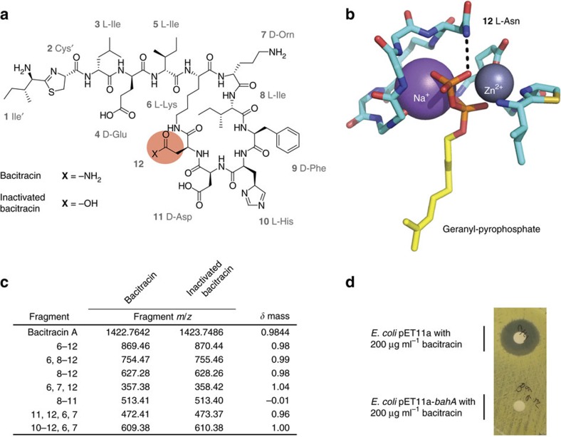
Figure 2. Paenibacillus sp. LC231 inactivates bacitracin through amidohydrolysis.
(a) Structure of bacitracin highlighting asparagine-12. (b) Structure of bacitracin A bound to geranyl-pyrophosphate (PDB 4K7T). Hydrogen bonding between bacitracin and the pyrophosphate moiety is indicated with dashed lines. (c) Molecular fragments of bacitracin and inactive bacitracin (R12->D12) identified using tandem mass spectrometry. The observed fragments correspond to the ring portion of the structure. Residues in each fragment correspond to the numbering in a. (d) Bacitracin inactivation by E. coli BL21(DE3) pET11a-bahA assayed using a Kirby–Bauer assay.

