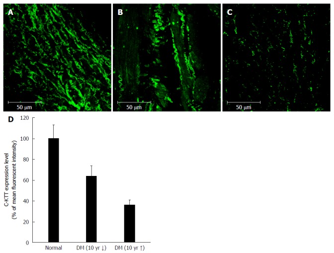
Figure 4.

Immunofluorescent staining of interstitial cells of Cajal in the human gastric corpus. Panels (A), (B), and (C) show representative images from the control, DM of < 10 years’ duration, and DM of > 10 years’ groups, respectively. Cellularity of ICC decreases with increasing duration of DM (D). DM: Diabetes mellitus; ICC: Interstitial cells of Cajal.
