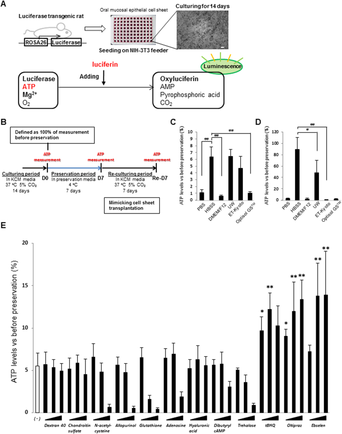
Figure 1. Screening system for determining the optimal preservation medium using oral mucosal epithelial cell (OEC) sheets derived from luciferase transgenic (luc tg) rats.
(A) Luminescence was induced in oral mucosal epithelial cell sheets derived from luc tg rats by adding luciferin. The observed luminescence correlates with the ATP levels. Scale bars represent 100 μm. (B) Time scale for ATP measurements using OEC sheets derived from luc tg rats. ATP levels of the cell sheet were measured at day 0 (D0, before preservation), day 7 ((D7) after preservation), and at 7 days after re-culturing (Re-D7). (C,D) Screening of basal medium using OEC sheets derived from luc tg rats. ATP levels at (C) day 7 after preservation and (D) re-culturing were calculated as the ration (%) of the ATP levels measured before preservation. *P < 0.05, **P < 0.001 (versus HBSS). Data are presented as mean ± SD of three independent experiments. (E) Screening of HBSS + additives using OEC sheets derived from luc tg rats. ATP levels at day 7 after preservation were calculated as the ration (%) of the ATP levels measured before preservation. *P < 0.05, **P < 0.001 (versus HBSS). Data are presented as the mean ± SD of three independent experiments.

