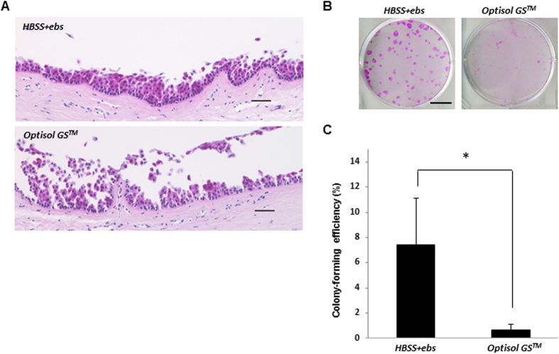
Figure 8. Morphology and colony-forming assay of human corneal limbal tissue after preservation in HBSS + ebselen or in Optisol GSTM.
(A) Hematoxylin and eosin staining of human corneal limbal tissues preserved in Optisol GS™ or HBSS + ebselen. Scale bars represent 50 μm. (B,C) Colony-forming assay of human corneal limbal tissues preserved in Optisol GS™ or HBSS + ebselen. Representative images of colonies in corneal limbal tissues after preservation (B). Scale bars represent 15 mm. *P < 0.05 was considered significant. Data represent the mean ± SD of four independent experiments (C).

