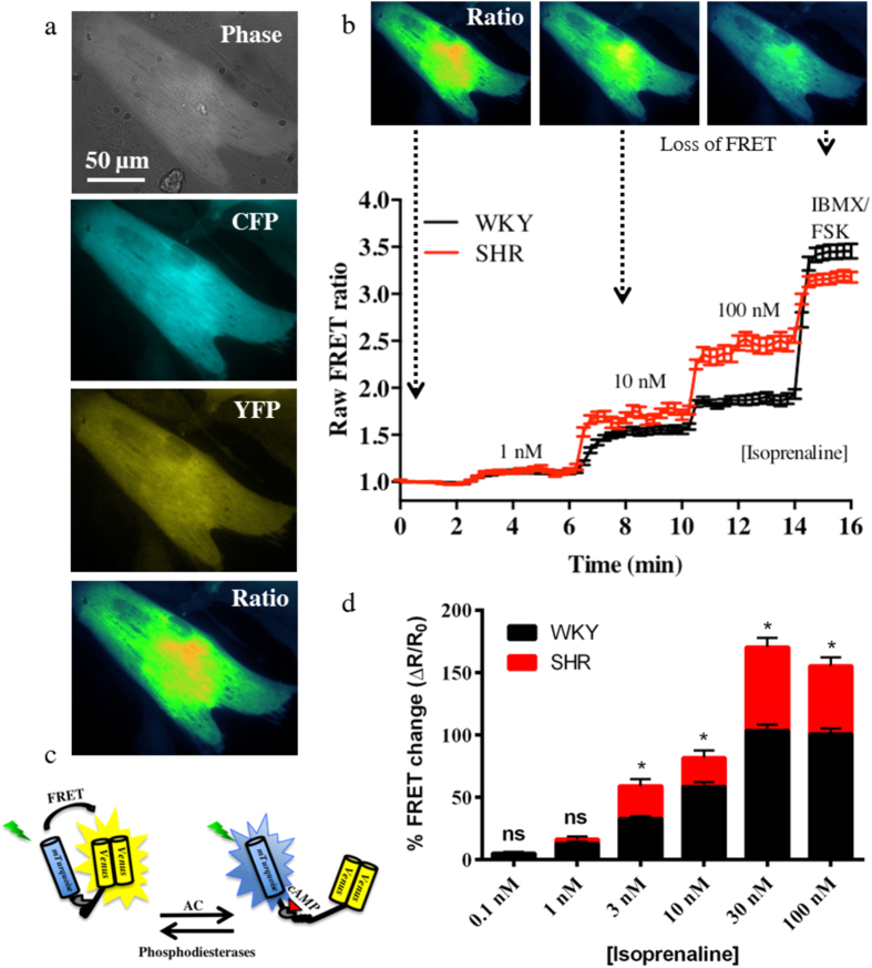
Figure 2. The ventricular myocytes from the pro-hypertensive SHR are hyper-responsive to β-adrenergic stimulation.
To investigate the β-adrenergic responses of the myocytes, we perfused the cells with isoprenaline and used a fluorescence resonance energy transfer (FRET) sensor Ad-Epac-SH187 to detect changes in cAMP levels as a result of the stimulation. CFP and YFP fluorescence intensities were taken and the ratio plotted (a). The binding of cAMP to Ad-Epac-SH187 (c) causes a conformational change in the sensor that moves the fluorophores apart, thereby resulting in loss of FRET (b). The SHR myocytes had significantly larger cAMP responses to isoprenaline when compared to the WKY at concentrations ≥ 3 nM (d). Concentrations and n numbers (WKY:animals/coverslip/cells and SHR:animals/coverslips/cells): 0.1, 3, 30 nM (WKY:28/12/55 and SHR:7/6/33), 1, 10,100 nM (WKY:28/11/50 and SHR:7/5/27). *p < 0.001, two-way ANOVA with Bonferroni correction. The cartoon in panel c was modified from Methods Mol. Biol. Simultaneous assessment of cAMP signaling events in different cellular compartments using FRET-based reporters, 1294, 2015, pp 1–12, Burdyga, A. & Lefkimmiatis, K. with permission of Springer60.

