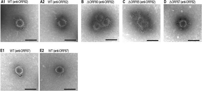
Figure 4. Immunoelectron microscopic analysis of the WT and mutant Sp5 phages.
Immunoelectron microscopic analyses were performed using the anti-ORF62 (A–D) or anti-ORF67 antibodies (E). WT and mutant Sp5 phages were initially treated with primary antibodies and then treated with secondary antibody-conjugated gold particles. Bar, 100 nm.

