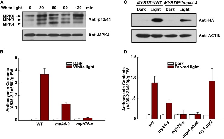
Figure 9.
MPK4 Is Involved in Light-Induced Anthocyanin Accumulation.
(A) Weak white light-induced MAPK activation. Four-day-old dark-grown wild-type seedlings were exposed to weak white light (40 μmol m−2 s−1) for the indicated times. MAPK activity was analyzed by immunoblotting with Phospho-p44/42 MAPK (Erk1/2) antibody (top panel), and MPK4 protein was determined by immunoblotting (bottom panel).
(B) Anthocyanin contents of 8-d-old seedlings of the wild type, mpk4-3, and myb75-c grown on plates under continuous dark or weak white light (40 μmol m−2 s−1). FW, fresh weight. Error bars represent sd of three replicates (as described in Figure 2B).
(C) Protein levels of MYB75 in the wild type and mpk4-3 grown under weak white light. 35S:MYB75WT/WT and 35S:MYB75WT/mpk4-3 were segregated from mpk4-3 heterozygous plants. Eight-day-old seedlings grown on plates under weak white light were dark-adapted for 4 d and then placed in the dark or continuous weak white light for another 9 h. The levels of MYB75 were determined by immunoblotting with anti-HA antibody.
(D) Anthocyanin contents of 8-d-old seedlings of the wild type, mpk4-3, myb75-c, phyA phyB, and cry1 cry2 grown on plates under continuous dark and far-red light (10 μmol m−2 s−1). FW, fresh weight. Error bars represent sd of three replicates (as described in Figure 2B).

