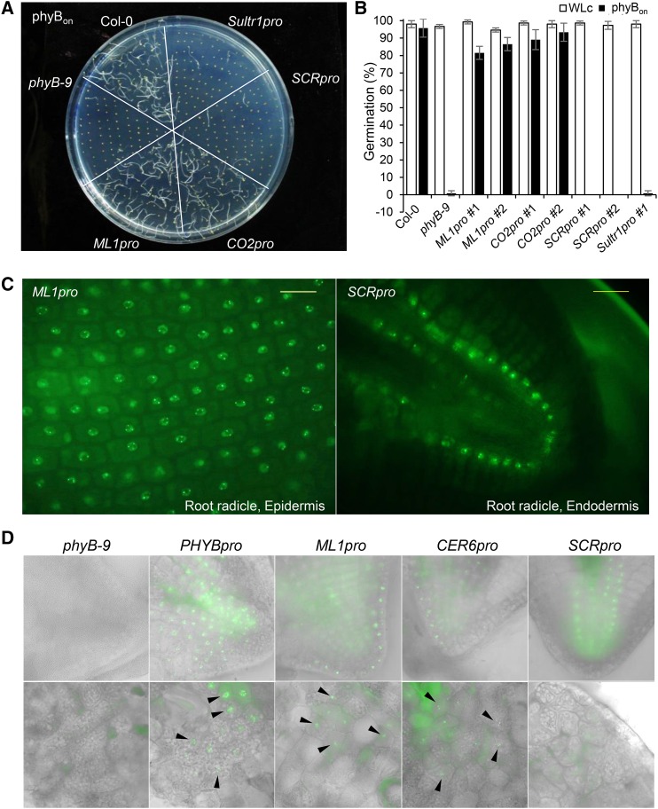
Figure 2.
Epidermal phyB Promotes Seed Germination in Response to Red Light.
(A) Visualization of germination frequencies for tissue-specific phyB lines under conditions that activate phyB. Seeds were surface-sterilized, treated with far-red light (2.56 μmol m−2 s−1) for 5 min followed by red light (11.4 μmol m−2 s−1) for 5 min, and then incubated in the dark for 4 d (phyBon).
(B) Germination frequencies for each tissue-specific phyB line. White bars indicate the continuous white light (WLc) condition, and black bars indicate the phyBon condition. Error bars indicate sd (n = 3 biological replicates, 50 seeds each).
(C) Epifluorescence images showing the expression of phyB-GFP in the epidermis (ML1pro) and endodermis (SCRpro) of imbibed seeds. Seeds were imbibed for 21 h before their seed coats were removed for epifluorescence imaging. Bars = 20 μm.
(D) Epifluorescence images showing the expression of phyB-GFP in the endosperm of imbibed seeds. Seeds were imbibed for 21 h before their seed coats were removed for epifluorescence imaging. Bright-field images in gray scale were merged with the GFP channel. The black arrowheads indicate phyB-GFP in the endosperm.

