Figure 6.
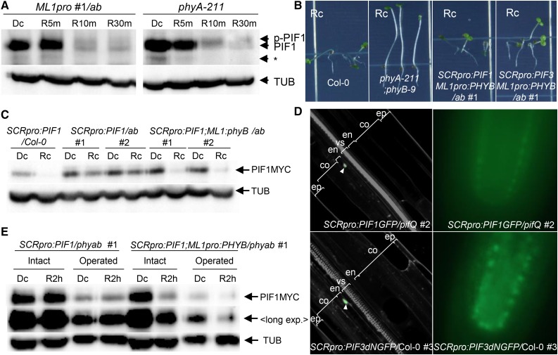
Epidermal phyB Non-Cell-Autonomously Promotes the Degradation of Endodermal PIFs.
(A) Epidermal phyB induces endogenous PIF1 degradation. Dark-grown seedlings were transferred to red light (9 μmol m−2 s−1) for 5, 10, or 30 min (R5m, R10m, or R30m) before being sampled to measure PIF1 levels using an anti-PIF1 antibody. PIF1 indicates endogenous PIF1, while p-PIF1 indicates the slower-migrating phosphorylated form. TUB indicates α-tubulin. The asterisk indicates nonspecific bands.
(B) Epidermal phyB inhibits hypocotyl negative gravitropism in lines expressing PIFs specifically in the endodermis. These images were taken after allowing the seedlings to grow on vertical plates for 4 d in continuous red light (Rc).
(C) Epidermal phyB induces degradation of endodermal PIF1. Seedlings were grown either in the dark (Dc) or in continuous red light (Rc) for 4 d before detection of MYC-tagged endodermal PIF1 (PIF1MYC) with an anti-MYC antibody.
(D) Confocal (left) and epifluorescence (right) microscopic images showing endodermis-specific GFP signal (green; white arrowheads) of 3-d-old dark-grown SCRpro:PIF3dNGFP/Col-0 seedlings counterstained with PI (gray). PIF3dN lacks the N-terminal 300 amino acids of wild-type PIF3. ep, epidermis; co, cortex; en, endodermis; vs, vascular structure.
(E) Epidermal phyB induces degradation of endodermal PIF1 in seedlings lacking cotyledons and the shoot apex. Four-day-old dark-grown seedlings were subjected to cotyledon and shoot apex removal (Operated) under a green safety light before being transferred back to either the dark (Dc) or to red light for 2 h (R2 h). MYC-tagged PIF1 was detected using an anti-MYC antibody. “Long exp.” indicates higher exposure.

