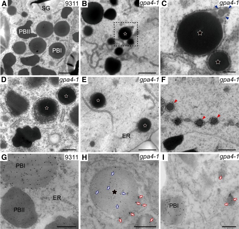
Figure 3.
Ultrastructure of Subaleurone Cells of Developing Endosperm of the Wild Type and gpa4-1 Mutant.
(A) Two types of protein bodies were observed in wild-type endosperm. Bar = 2 μm.
(B) The first type of novel structure was observed from 9 DAF in endosperm of the gpa4-1 mutant. Stars represent this type of novel structure in (B) to (E). Bar = 1 μm.
(C) Enlarged image of the boxed area in (B). Triangles indicate protein aggregates in the periphery of the first type of novel structure. Bar = 200 nm.
(D) The first type of novel structure in 12 DAF endosperm cells. Bar = 1 μm.
(E) The first type of novel structure is directly linked with the ER. Bar = 1 μm.
(F) The second type of novel structure (red triangles) in 12 DAF endosperm cells. Bar = 500 nm.
(G) to (I) Immunoelectron microscopy localization of glutelins and prolamins in subaleurone cells of developing wild-type (G) and the gpa4-1 mutant ([H] and [I]) endosperm. Bars = 500 nm in (G) and (H) and 200 nm in (I).
(G) Glutelins and prolamins are accumulated separately in the wild type.
(H) The first type of novel structure contains a glutelin (6-nm gold, blue arrows) core and a prolamin (15-nm gold, red arrows) periphery.
(I) The second type of novel structure contains prolamins (15-nm gold, red arrows).

