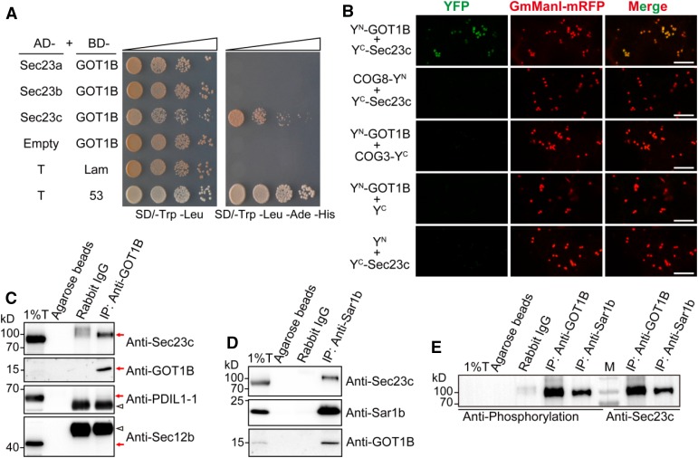
Figure 7.
GOT1B Physically Interacts with Sec23 Proteins.
(A) Y2H assay showing that GOT1B interacts with Sec23c. The bait plasmid (pGBKT7-Lam or pGBKT7-53) was cotransformed into the AH109 yeast strain with the prey plasmid (pGADT7-T) to serve as negative and positive controls, respectively.
(B) BiFC assay showing that GOT1B can interact with three Sec23 isoforms in leaf epidermal cells of N. benthamiana. The Golgi-localized membrane proteins COG3 and COG8 (a pair of interacting proteins) were used as the negative control (Tan et al., 2016). Bars = 10 μm.
(C) Co-IP assay showing that Sec23c can be immunoprecipitated by anti-GOT1B polyclonal antibodies in total extract of developing endosperm. Immunoblots with anti-PDIL1-1 and anti-Sec12b antibodies showing no ER lumenal and membrane protein contamination in the IP samples. Red arrows indicate the corresponding protein bands. Triangles indicate the heavy chain of rabbit IgG protein. T, total extract; IP, immunoprecipitation; IB, immunoblot.
(D) Co-IP assay showing that both Sec23c and GOT1B can be immunoprecipitated by anti-Sar1b antibodies in total extract of developing endosperm.
(E) Immunoblot analysis of the IP samples with antiphosphorylation antibodies (left) and anti-Sec23c antibodies (right), respectively. M, protein markers.

