Figure 1.
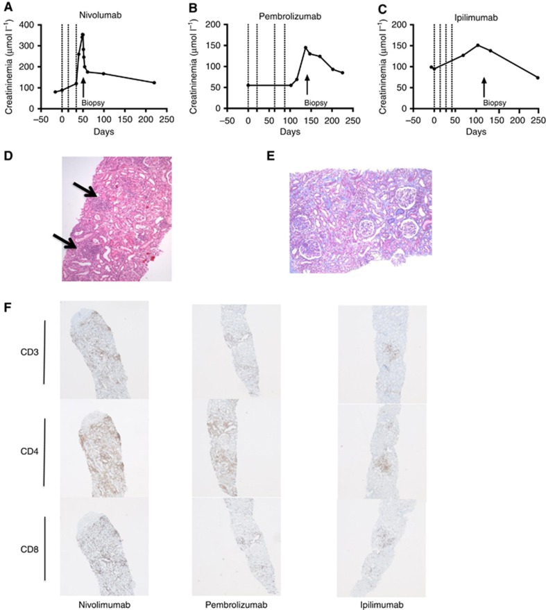
Interstitial nephritis following the use of immune checkpoint inhibitors. Serum creatinine is depicted according to the time that patients were treated with nivolumab (A), pembrolizumab (B), or ipilimumab (C). Each vertical line indicates one injection of an immune checkpoint inhibitor. The arrow represents a kidney biopsy. (D and E) Hematoxylin-eosin and Masson' trichrome staining of a kidney section (original magnification) revealed acute interstitial nephritis with lymphoplasmacytic cell infiltrate and acute tubular injuries. Arrows indicate granulomatosis aspect of the infiltrate. (F) Anti-CD3, CD4, and CD8 staining (original magnification x50). The interstitial inflammatory infiltrate was mainly positive for the CD3 CD4 T-cell marker. Anti-perforin, granzyme, and CD56 were negative (not shown).

