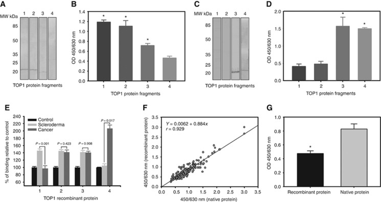
Figure 5.
Identification of the novel TAA as a fragment of human DNA-topoisomerase I. (A) Western blot analysis of the anti-SCL-70 autoantibody reactions with four recombinant fragments of TOP1. (B) ELISA analysis of the anti-SCL-70 autoantibody reactions with four recombinant fragments of TOP1. The results represent the mean+s.e. for each group (*P<0.001). (C) Western blot analysis of the rabbit polyclonal antibody reactions with four recombinant fragments of TOP1. (D) ELISA analysis of the rabbit polyclonal antibody reactions with four recombinant fragments of TOP1. The results represent the mean+s.e. for each group (*P<0.001). (E) Comparison of serum immune reaction to recombinant TOP1 fragments among cancer patients, scleroderma patients and healthy donors. The immune reactions in the serums of the cancer patients and scleroderma patients are presented as percentages of mean+s.e. relative to the serums of the healthy controls for each fragment. The P-values indicate the comparisons of the immune reactions in serums between the cancer patients and the scleroderma patients to the each recombinant fragment. (F) One hundred and eighty serum (180) samples were collected from cancer patients, healthy donors and other non-cancer patients. Serum autoantibody against the recombinant TOP1 amino acid sequence 329–765 fragment and the chromatographically purified novel TAA were determined simultaneously, with the correlation being analysed using a linear regression technique. A strong correlation was observed (y=0.00062+0.844X, r=0.929, P<0.0001). (G) Comparison of antibody affinity of the rabbit anti-novel TAA antibody to the recombinant TOP1 amino acid sequence 329–765 fragment and the chromatographically purified novel TAA. The results represent the mean+s.e. for each group (*P<0.001).

