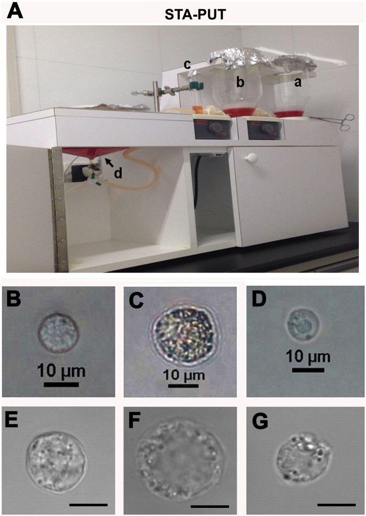
Figure 1. Isolation and morphological characteristics of human spermatogonia, pachytene spermatocytes and round spermatids.
(A), STA-PUT apparatus was utilized to separate human spermatogonia, pachytene spermatocytes and round spermatids using 4%, 2% and 0.5% BSA. Notes: a, gradient glass chamber with 4% BSA; b, gradient glass chamber with 2% BSA; c, cell loading chamber with 0.5% BSA; d, standard sedimentation chamber. (B–D), Phase-contrast microscope showed the morphological characteristics of the freshly isolated human spermatogonia (B), pachytene spermatocytes (C) and round spermatids (D). (E–G), DIC microscope revealed the appearance of the freshly isolated human spermatogonia (E), pachytene spermatocytes (F) and round spermatids (G). Scale bars in E, F and G = 10 μm. The data shown in (B–G) were representatives from eight independent experiments of thirty patients.

