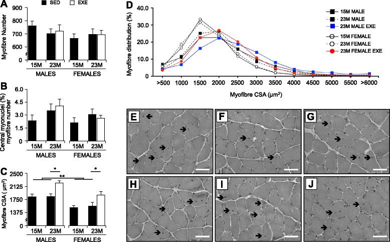
Fig. 3.

Morphometric characterization of the soleus muscles from 15-months SED, 23-month SED, and 23-month RWE mice, of both sexes. Entire transverse sections of the soleus muscles stained with H&E were used to quantify the number of individual muscle fibers (a) and proportion of myofibers with non-peripheral nuclei or central nucleation (b). Average myofiber size (as measured by cross-sectional area (CSA)) (c) and myofiber size distribution (d) were quantified on transverse section of the soleus immunostained for laminin. Myofibers with displaced or central nuclei were seen in the soleus muscles of male (e–g) and female (h–j) mice at both 15-month SED (e, h) and 23-month SED (f, i), as well as after RWE (g, j) (arrows). Data were analyzed by ANOVA, using age and sex and sex and activity as variables. Data are mean ± SEM. Asterisk denotes significance at *P < 0.05; **P < 0.01; ***P < 0.001. For each age group, N = 6–10 mice/group. Scale is 50 μm
