Fig. 5.

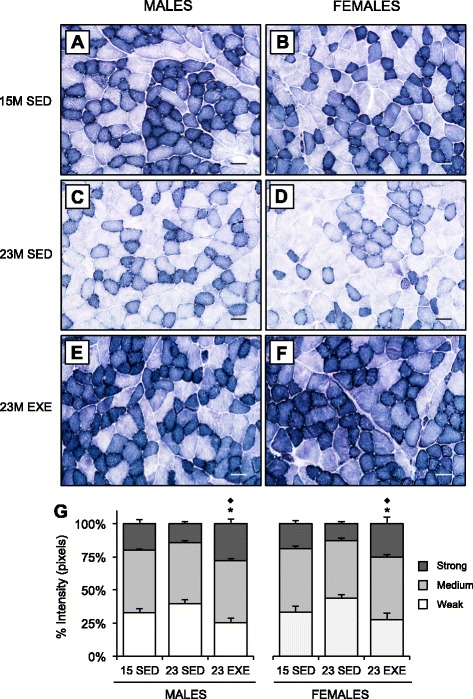
NADH-TR staining intensity in the quadriceps muscles of 15-month SED, 23-month SED, and 23-month RWE mice, of both sexes. Transverse sections of the quadriceps muscles stained with NADH-TR were used to quantify changes to oxidative state. Representative images were taken at ×20 magnification in male (a, c, e) and female (b, d, f) mice from both 15-month SED (a, b) and 23-month SED (c, d) cohorts, as well as after RWE (e, f) in the rectus femoris. Scale is 50 μm. NADH-TR intensity was quantified by calculating the percentage of weak, intermediate, or strong staining intensity of the whole muscle relative to total pixel intensity (g). Data for each intensity level was analyzed by ANOVA, using age and sex and sex and activity as variables. Asterisks indicate a significant decrease in the percentage of weakly staining intensity and diamond an increase in the percentage of strongly staining intensity between 23-month SED and 23-month EXE cohorts of the same sex (P < 0.05). Data are mean ± SEM. For each age group, N = 6–9 mice/group
