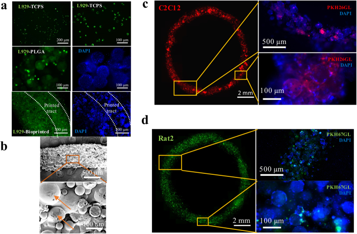
Figure 7. Characterisation of bioprinted ring constructs.
(a) Fluorescence images of L929 cells labelled by green fluorescent cell linker PKH67GL cultured on TCPS, PLGA microspheres, and printed construct. High cell density could be observed on the printed construct. (b) SEM images of the printed construct using L929 cells. The construct was fully covered with cells. Obvious cells are indicated by arrows. Fluorescence images of printed constructs using (c) C2C12 cells labelled with red fluorescent cell linker PKH26GL and (d) Rat2 cells labelled by green fluorescent cell linker PKH67GL after 3 days of culture. The fluorescent signals proved that the cells are still viable. Blue fluorescence images (right) in (a,c,d) show DAPI (nuclei) staining of the corresponding samples.

