
Figure 2. Predicted Lipid Binding Pocket of Prohibitin.
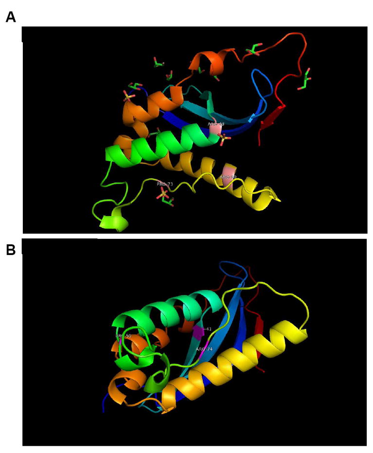
Based on sequence alignment, a 3D model of prohibitin was built using the 3D structure 2DYB chain ‘A’ as template. (A) 2DYB chain A is the Protein Databank structure for p47 Phox and it is based on the putative binding pocket residues that alignment with amino acids in the prohibitin sequence that can serve as a binding pocket in prohibitin-PIP3 binding (B). The p47 Phox template shares 17% identities with the query sequence using the ALIGN program.