
Figure 5.
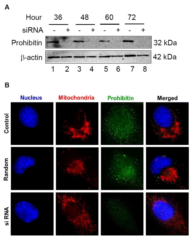
A. Downregulated prohibitin is shown by siRNA knockdown.
ARPE-19 cells were transfected by prohibitin specific siRNA in serum free medium in a time-dependent manner (36 to 72 h). Prohibitin expression and depleted levels were analyzed by Western blotting analysis from cell extracts. Prohibitin levels were diminished during siRNA knockdown. β-actin was used as a loading control.
B. Mitochondrial morphological changes during prohibitin siRNA knockdown analysis. ARPE-19 cells were incubated using prohibitin specific siRNA (175 ng for 48 h) and random sequence control. Prohibitin and organelles were visualized by immunocytochemical analysis using DAPI (blue, nucleus), MitoTracker Orange (red, mitochondria), and Alexa-Fluor 488 (green, prohibitin). Disrupted mitochondrial morphological changes were observed under prohibitin depleted levels. The scale bar represents 5 μm.
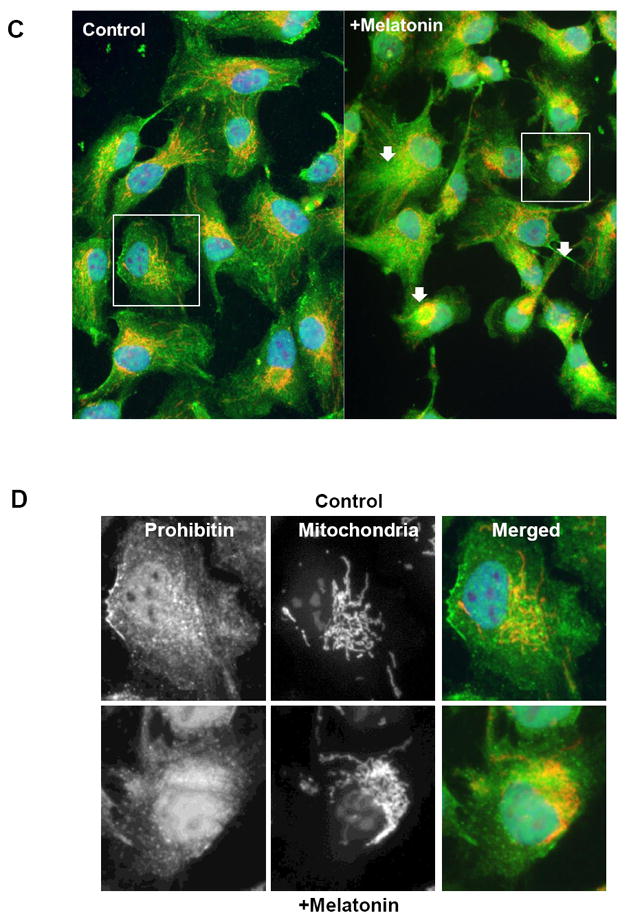
C, D. ARPE19 cells were incubated using 100μM DMSO-dissolved melatonin for 30 mintures, followed by incubation in untreated medium (12 hours). Cells were visualized using prohibitin antibody and Alexa-Fluor-488 secondary antibody. Mitochondria and nucleus were labeled using MitoTracker Orange and DAPI respectively. Cell morphology was tracked at initial, half-hour, and 12-hour time points. Cells incubated for 12 hours are shown.