
Figure 6.
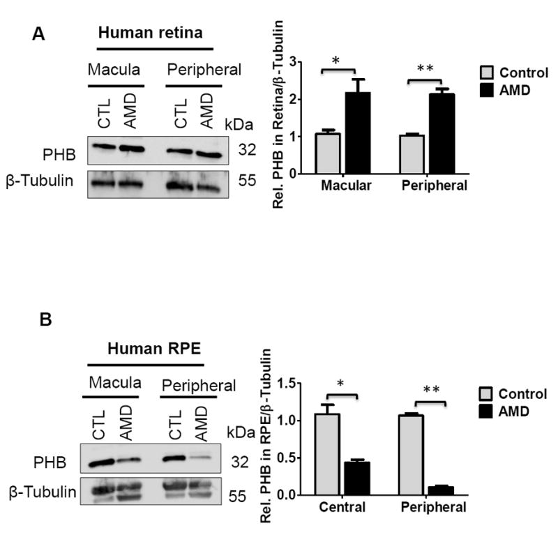
Prohibitin expressions in human AMD retina (8mm macular, peripheral retina) and RPE (8mm central and peripheral region, n=6 for two biological sample for triplicate experiments)) were analyzed by Western blotting. Retinal tissue and RPE cells were homogenized in RIPA buffer followed by sonication. As a loading control, β-tubulin was used. Prohibitin was analyzed quantitatively based on pixel size and intensity. A. Prohibitin in the macular and peripheral region from AMD retina. B. Prohibitin in the central and peripheral region from AMD RPE. Statistical comparisons between means were performed by 2-tailed t test. A p value of ≤0.05 was considered as statistically significant (P > 0.05 not significant; P ≤ 0.05 *; P ≤ 0.01 **; P 0.001 ***).