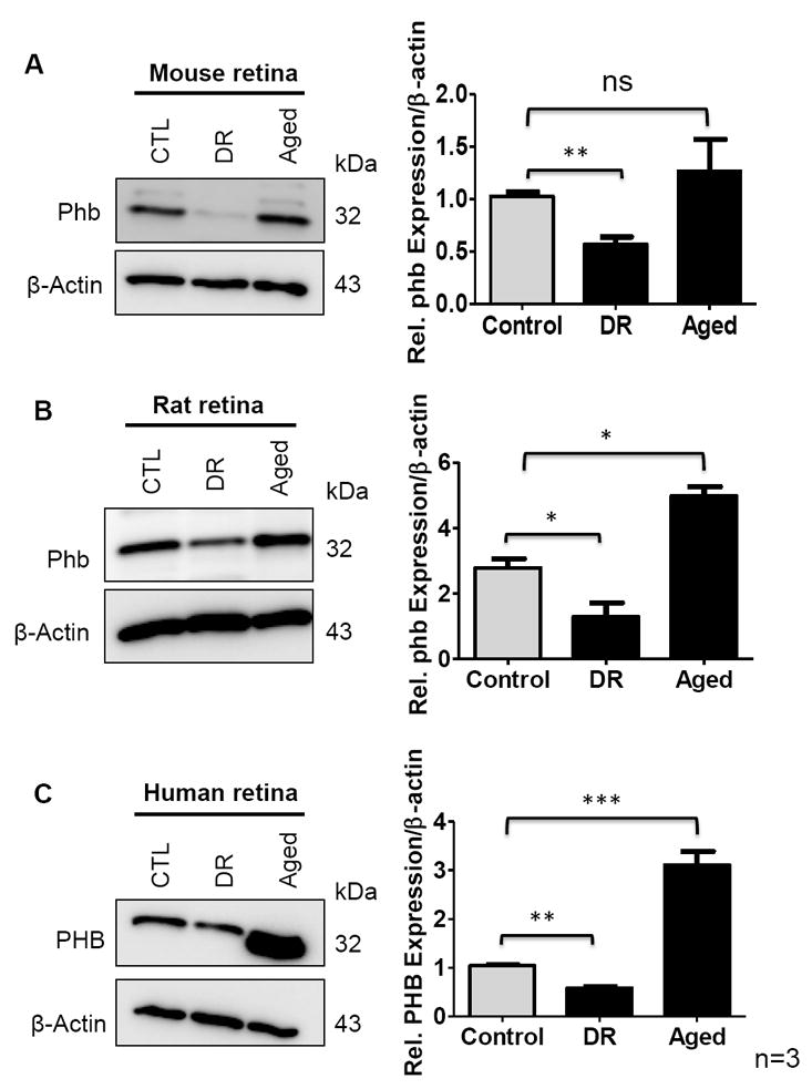
Figure 7.

Prohibitin in control, diabetic retinopathy (DR) and aged retina (n=9 for biological triplicate and technical triplicate) were analyzed. Retinal tissue was homogenized in RIPA buffer followed by sonication. β-actin blot was used as a loading control. A. Prohibitin from diabetic retinopathy (DR) and aged mouse retina was compared to control. B. Prohibitin from diabetic retinopathy (DR) and aged rat retina was compared to control. C. Prohibitin from diabetic retinopathy (DR) and aged human retina was compared to control. Statistical comparisons between means were performed by 2-tailed t test. A p value of ≤ 0.05 was considered as statistically significant (P > 0.05 ns (not significant); P ≤ 0.05 *; P ≤ 0.01 **; P ≤ 0.001 ***).
