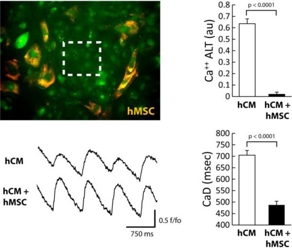
Figure 2.
Image of hCM monolayer co-cultured directly with hMSC (orange). Ca++ transients were recorded from a region indicated by the white box. The effects of hMSC (10000 cells) on hCM under conditions of oxidative stress are shown. Ca++ transient recordings in the presence of H2O2 (left) show a decrease in Ca++ ALT with hMSC compared to hCM alone. The graphs (right) show summary data for hCM alone or with hMSC. Overall, hMSC significantly decreased Ca++ ALT (n=9, p< 0.0001) and CaD (n=8, p<0.0001).

