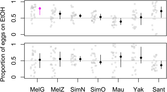
Fig. 2.

Oviposition on 6% ethanol vs 0% ethanol. Each strain was presented with the two substrates touching (a split-patch, top figure) or separated by 7 cm (two patches, bottom figure). Each replicate is shown as a gray circle, and the median for each strain is shown as a filled circle with a line spanning the central 50% range. Strains significantly different than random based on a binomial test, Bonferroni corrected for seven parallel tests, are shown in fuchsia.
