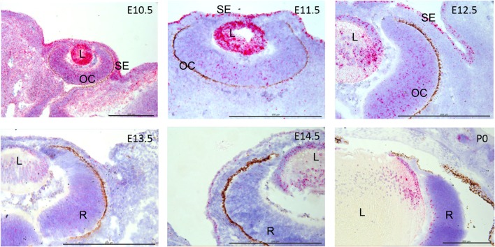
Fig. 3.
Adamts18 mRNA localization during mouse eye development. Adamts18 mRNA, indicated by red dots overlying cells, is strongly expressed in the lens (L) throughout the development of the eye, with strongest expression at E10.5 and E11.5 and declining thereafter. At E10.5, E11.5, and E12.5, Adamts18 mRNA is also expressed in the surface ectoderm (SE) and the optic cup (OC). At E13.5, E14.5, and at birth (P0), Adamts18 mRNA is localized to lens nuclei and lens epithelium. At P0, Adamts18 mRNA is expressed in the innermost layer of the developing retina (R), and the ganglion cell layer. Hematoxylin counterstain is blue. Scale bars: 200 µm.

