Fig. 4.

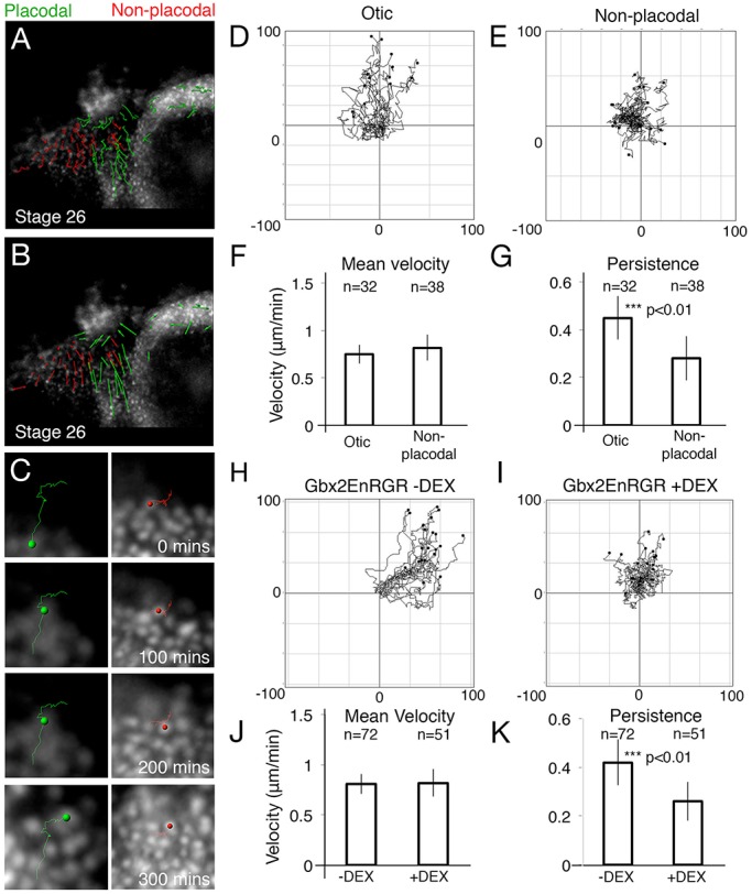
Gbx2 targets are required for directional cell movements into the otic placode. (A,B) Automatic tracks of PPR cells sorted into placodal (green) or non-placodal (red) cells based on final position and overlaid onto the final frame of the movie (A). (B) Cell displacements, arrows indicate the displacement of cells from their initial to final positions. (C) An example of five cells that were automatically tracked in a region posterior and ventral to the forming otic placode. (D,E) Summary diagrams of all tracked cells. To compare the directionality of cells, tracks were translated as in Fig. 3. (F,G) Student t-tests (2-tailed) of otic, epibranchial (epi) and non-placodal cell movements to analyse mean velocity (F; P=0.18) and persistence (G; P=2.5×10⁻⁴). (H,I) Cells were tracked from PPR grafts containing Gbx2-EnR-GR in the absence (H) and presence (I) of DEX from stage 18. (J,K) Students t-tests (2-tailed) of cells carrying Gbx2-EnR-GR in the presence or absence of DEX to analyse mean velocity (J; P=0.51) and persistence (K; P=1.5×10⁻³) of cell movements. n=number of cells tracked from three independent movies. Error bars: ±one standard deviation either side of the mean.
