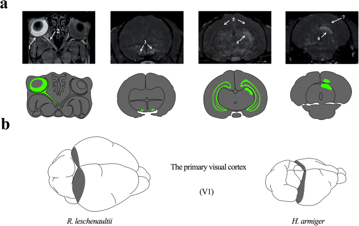
Figure 1.
MEMRI of R. leschenaultii and the location of V1 in bats. a). MRI of the Mn2+-enhanced visual pathway performed 24 hours after the intravitreal injection of Mn2+ into the left eye of R. leschenaultii. Annotations: retina (1), optic nerve (2), optic chiasm (3), lateral geniculate nucleus (4), hippocampus (5), superior colliculus (6) and primary visual cortex (7). b). The primary visual cortices of R. leschenaultii and H. armiger.

