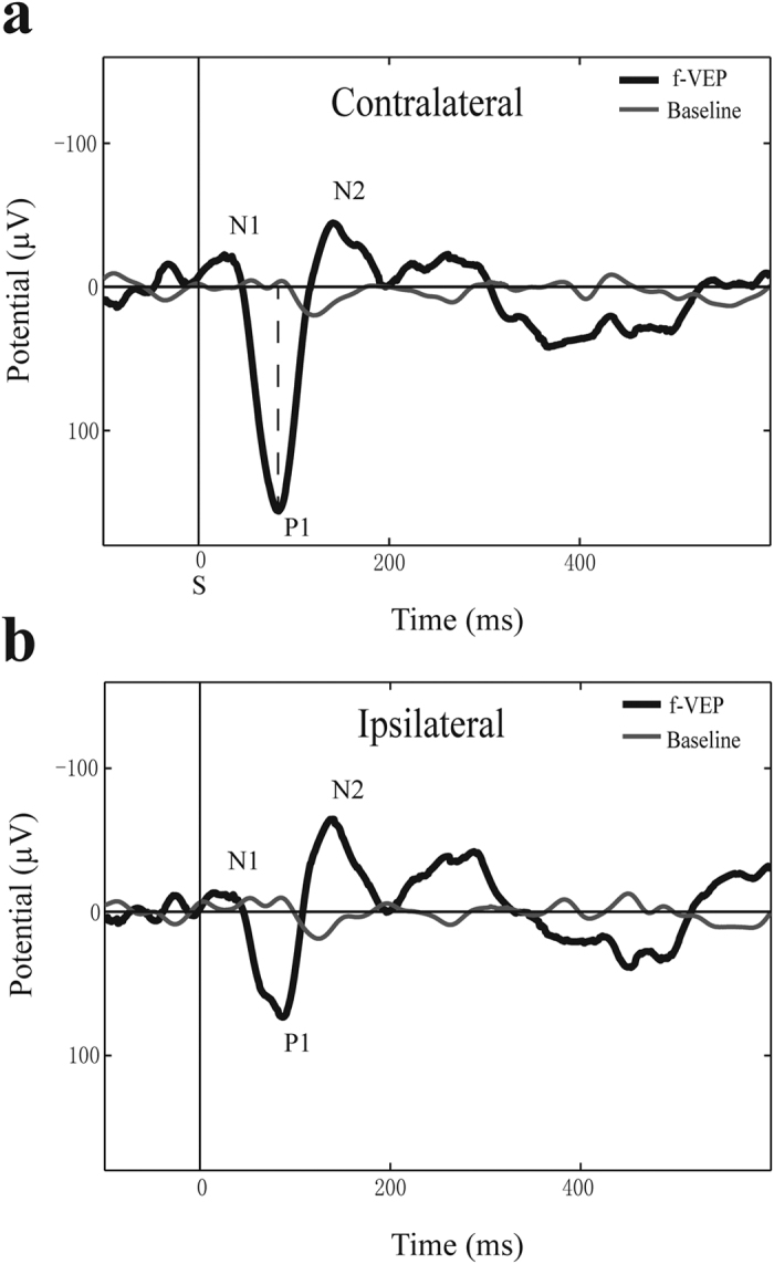
Figure 3.

Representative flash VEP waveforms from C. sphinx. a). The contralateral cortex f-VEP. b). The ipsilateral cortex f-VEP. Annotations: the N1-P1-N2 peak structure; vertical dashed line represents the position of the P1 amplitude measure; S indicates flash stimulus.
