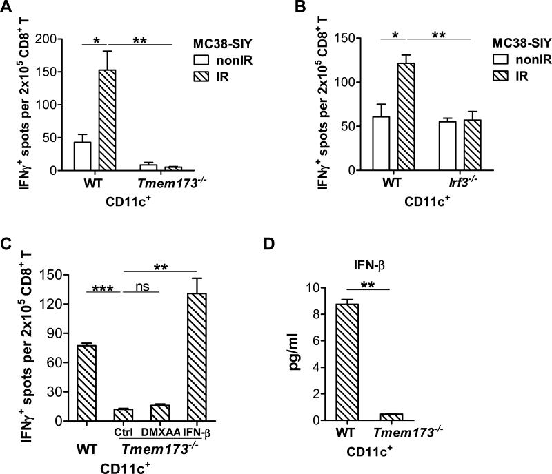
Figure 3. STING-IRF3 axis in DCs is activated by irradiated-tumor cells.
(A-C) BMDCs were cultured with 40Gy-pretreated MC38-SIY cells or non-irradiated-MC38-SIY cells. Subsequently purified CD11c+ cells were co-cultured with isolated CD8+ T cells from naive 2C mice for three days and analyzed by ELISPOT assays. (A) BMDCs from WT or Tmem173−/− mice were used for co-culture with irradiated or non-irradiated MC38-SIY cells. DC cross-priming activity was analyzed by ELISPOT assays. (B) BMDCs from WT or Irf3−/− mice were used for co-culture with irradiated or nonirradiated MC38-SIY cells. DC cross-priming activity was analyzed by ELISPOT assays. (C) WT and Tmem173−/− BMDCs were cultured with 40Gy-pretreated MC38-SIYhi cells. 10ng/ml IFN-β was added into the co-culture of Tmem173−/− BMDC and irradiated-MC38-SIY cells. 100μg/ml DMXAA was added to isolated Tmem173−/− CD11c+ cells for additional three hours incubation. DC cross-priming activity was analyzed by ELISPOT assays. (D) WT and Tmem173−/− BMDCs were co-cultured with 40Gy-pretreated MC38-SIY cells. The purified CD11c+ cells were incubated for additional two days and the supernatants were collected to measure IFN-β by ELISA assay. STING-deficient mice are represented by Tmem173−/−. Representative data are shown from three (A-D) experiments. Data are represented as mean ± SEM. *P<0.05, **P < 0.01, ***P < 0.001 and ns No significant difference (Student's t test). See also Figure S2 and S3.

