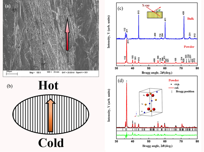
Figure 1.
(a) The SEM image of fractured DyNiSi button. (b) The schematic for the formation of texture structure in DyNiSi compound during arc-melting. (c) XRD patterns carried out on DyNiSi powder and on the bulk surface perpendicular to the longitudinal direction of columnar grains. (d) The observed (dots) and calculated intensities (line drawn through the data points) of powder XRD patterns at room temperature. The short vertical lines indicate the Bragg peak positions of TiNiSi-type orthorhombic structure. The lower curve shows the difference between the observed and calculated intensities. The inset shows the perspective view of the unit cell of DyNiSi.

