Figure 5.
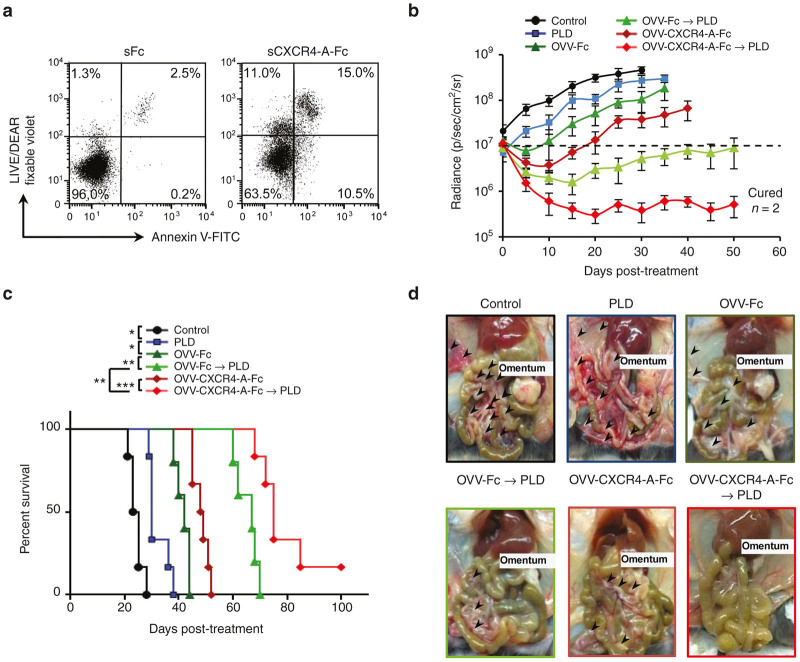
Effect of the CXCR4-A-Fc fusion protein on ID8-R tumor growth. (a) Cell death in ID8-R tumor cells treated with soluble CXCR4-A-Fc fusion protein (100 µg/ml) for 24 hours was determined by staining with Annexin V-FITC and LIVE/DEAD fixable violet. Tumor cells treated with soluble Fc fragment of mouse IgG2a serve as controls. One representative experiment of three independent experiments performed is shown. (b) C57BL/6 mice (n = 8 − 10) were injected i.p. with 2 × 10⁵ ID8-R cells. Oncolytic virotherapy with OVV-CXCR4-A-Fc or OVV-Fc (108 PFU delivered i.p.) was initiated 10 days later. In parallel experiments, tumor-bearing mice were treated with PLD (10 mg/kg) delivered i.v. or PLD was delivered to virally-treated mice 8 days after virus injection. Control mice were treated with PBS. Tumor progression was monitored by bioluminescence imaging using the Xenogen IVIS Imaging System. Data points represent mean ± SD. (c) Kaplan–Meier survival plots were prepared and significance was determined using the log-rank method. *P < 0.05, **P < 0.01, ***P < 0.001. (d) Metastatic dissemination in the omentum, diaphragm, mesentery and peritoneal wall was assessed by identifying metastatic colonies (>5 mm) in individual mice at the time of development of bloody ascites in control mice. Representative images of metastasis within the peritoneal cavity of one mouse from each group are shown. OVV, oncolytic vaccinia virus; PBS, phosphate buffered saline; PFU, plaque forming units; PLD, pegylated liposomal doxorubicin; SD, standard deviation.

