
Figure 1.
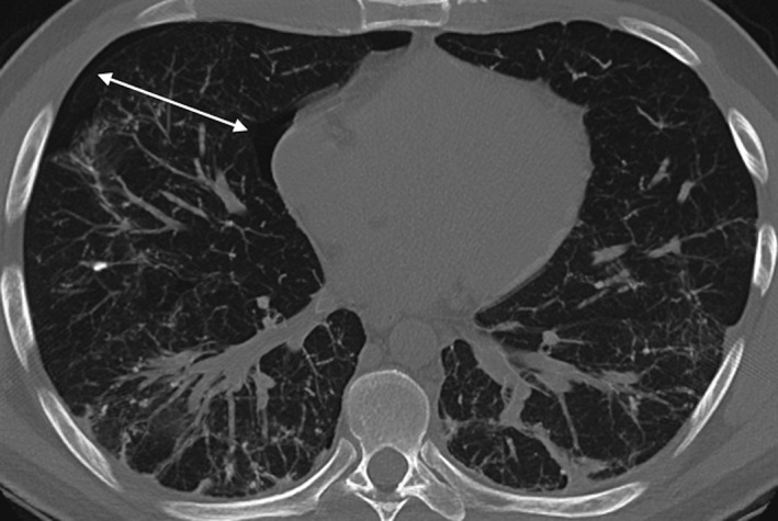
Thoracic computed tomography scan showing a small right‐sided pneumothorax (white arrows), with background subpleural and peribronchovascular opacities predominantly in the posterior dependent distribution areas. Dense partly calcified linear reticulations in these regions were most consistent with dendriform pulmonary ossification.