
Figure 2.
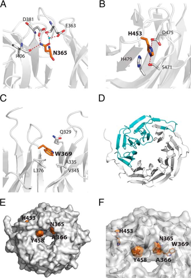
Schematic representations of the mutated amino-acids. The mutated amino-acids are shown on the crystal structure of the TBL1XR1 WD40 domain (PDB ID 4LG9). In each case the mutated residues are shown in orange, TBL1XR1 in gray, and water molecules in the crystal structure in cyan. The numbering of the amino acids is as for TBL1X. A, N365. B, H453. C, W369. D, c.1312-1G>A splice mutation with the missing amino acids in cyan (starting at asterisk). E, Surface representation of the WD40 domain to show the mutations that are on the surface. F, Transparent representation of the WD40 domain to show the buried and surface mutations.