Figure 3.
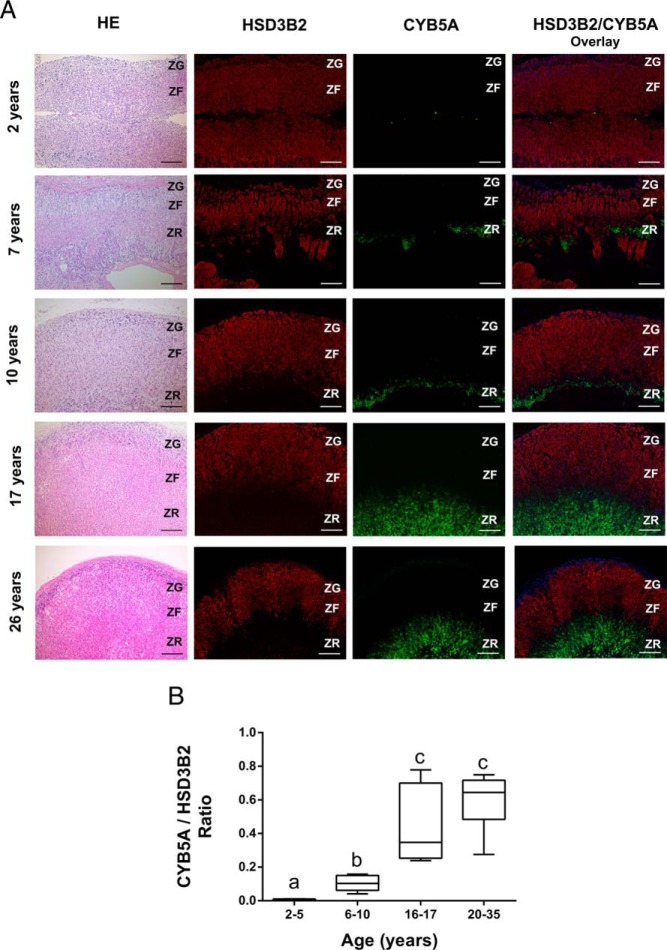
Immunolocalization of HSD3B2 and CYB5A in the human adrenal. A, Representative images of double immunofluorescence of HSD3B2 (red) and CYB5A (green) in adrenal glands from following age groups: 2–5 y (2 y male), 6–10 y (7 and 10 y males), 16–17 y (17 y female) and 20–35 y (26 y female). HSD3B2 and CYB5A are sharply segregated to the zona glomerulosa–ZF and the ZR, respectively. Nuclei are counterstained in DAPI (blue). Scale bars, 200 μm. B, Dual immunofluorescence was performed for HSD3B2 and CYB5A in the adrenal glands of the following age groups: 2–5 y (n = 4), 6–10 y (n = 6), 16–17 y (n = 6), and 20–35 y (n = 6). The areas expressing CYB5A (green) and HSD3B2 (red) were quantified using image analysis, and ratios of areas with CYB5A expression/HSD3B2 expression were then calculated and compared in different age groups. Data are represented as mean ± SEM. Statistical significance was determined using a one-way ANOVA followed by a Holm-Sidak test. Different letters above the bars indicate statistically significant differences (P < .05) between the different age groups.

