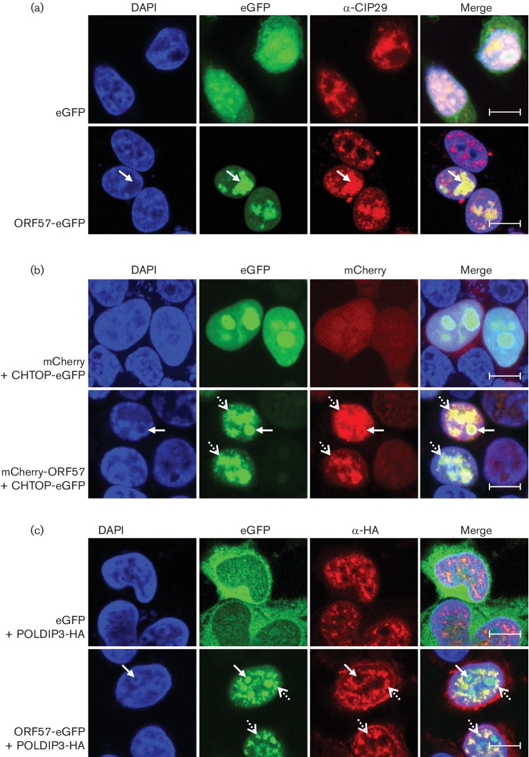
Fig. 2.
ORF57 recruits CHTOP and CIP29, but not POLDIP3, into the nucleolus. (a) Confocal microscopy of cells expressing eGFP or ORF57-eGFP. Cells were stained using a CIP29-specific antibody and the nucleus visualized using DAPI. Arrows indicate the nucleolus. Scale bar, 10 µm. (b) Confocal microscopy of cells expressing the indicated expression plasmids of ORF57 and CHTOP or POLDIP3, respectively. (c) Where appropriate, cells were stained using a HA-specific antibody and fixed using methanol, while all other cells were fixed using formaldehyde. The nucleus was visualized using DAPI. Arrows indicate the nucleolus, dashed arrows indicate nuclear speckles. Scale bar, 10 µm.

