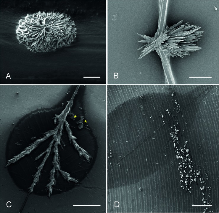
Fig 8.
In the scanning electron microscope the crystalin-like core of the normal thread droplet in A. luminosa appears A) as rosettes or B) sheet-shaped pointed bundle, when pre-dried in the desiccation chamber before attachment on stubs. C) Samples, attached to polished aluminum stubs and subsequently dried in the desiccation chamber; the crystals grow needle-shaped along the thread and form lateral branches. In addition to the crystal at the centre of the droplet, a non-crystalline content (yellow asterisks) could be observed at the base of the droplet. D) Samples, freshly attached to a riffled stub and air-dried; no crystal formation within the droplet area could be observed. Scale bar in A = 10 μm; B = 5 μm; C and D = 200 μm.

