
Fig 4. Example of immunostaining of different antibodies.
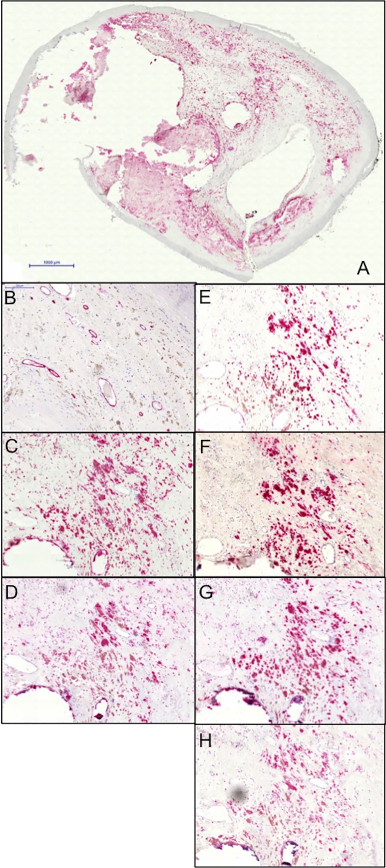
A: Overview. B: CD34 stained vessels. C: CD68 positive macrophages. D: HIF1α. E: VEGFR2. F: VEGF. G: NOTCH1. H: Dll4. Black bars represent 1000 μm (A) and 200 μm (B-H).
Official websites use .gov
A
.gov website belongs to an official
government organization in the United States.
Secure .gov websites use HTTPS
A lock (
) or https:// means you've safely
connected to the .gov website. Share sensitive
information only on official, secure websites.
A: Overview. B: CD34 stained vessels. C: CD68 positive macrophages. D: HIF1α. E: VEGFR2. F: VEGF. G: NOTCH1. H: Dll4. Black bars represent 1000 μm (A) and 200 μm (B-H).