
FIGURE 3:
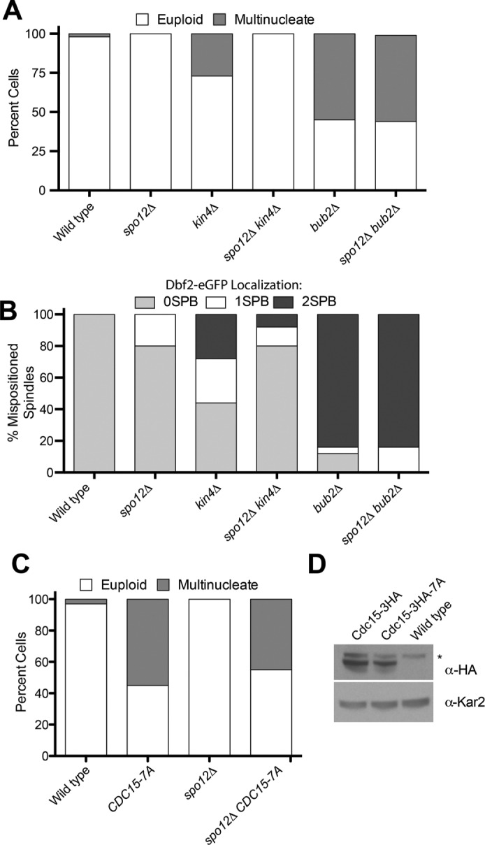
Inactivation of the FEAR network suppresses the SPoC defect of kin4Δ but not bub2Δ mutants. (A) Percentage multinucleate cells generated after one cell cycle. Cells were released from an α-factor pheromone–induced G1 arrest, and the percentage of multinucleate cells was assessed upon completion of the first cell cycle. Cells were scored as in Figure 2A. dyn1-AID kar9Δ (A35707), dyn1-AID kar9Δ spo12Δ (A35700), dyn1-AID kar9Δ kin4Δ (A35603), dyn1-AID kar9Δ spo12Δ kin4Δ (A36048), dyn1-AID kar9Δ bub2Δ (A36082), and dyn1-AID kar9Δ spo12Δ bub2Δ (A36965) cells harboring GFP-tagged α-tubulin were grown as described in Figure 2. Cells were released into the cell cycle in YEPD medium and then monitored by live-cell microscopy. Depletion of dyn1-AID was induced with 100 μM auxin in synthetic complete medium (n = 100). Note that dyn1-AID kar9Δ bubΔ (A36082) and dyn1-AID kar9Δ kin4Δ (A35603) are shown again from Figure 2A. (B) Dbf2-eGFP localization to SPBs in cells with mispositioned spindles in dyn1Δ (A36852), dyn1Δ spo12Δ (37134), dyn1Δ kin4Δ (A36782), dyn1Δ kin4Δ spo12Δ (A36851), dyn1Δ bub2Δ (A36850), and dyn1Δ spo12Δ bub2Δ (A36722) strains. Cells were incubated for 24 h at 14°C in YPD medium supplemented with 0.3 mM adenine hemisulfate, 0.4 mM l-tryptophan, 1 mM l-histidine, 0.2 mM uracil, and 1 mM l-leucine to induce spindle mispositioning. Cells were then fixed in 4% PFA and imaged. All strains expressed mCherry-tagged α-tubulin to mark the spindle. (C) Percentage of multinucleated and euploid cells was assessed in CDC15 (A38964), CDC15-7A (A38963), spo12Δ CDC15 (A38294), and spo12Δ CDC15-7A (A38290) strains that had mispositioned their spindles. Spindle mispositioning was induced as in Figure 2, and cells were analyzed as in Figure 2A. (D) Western blot analysis of whole-cell lysates to examine the expression of Cdc15-HA3 (A38294), Cdc15-HA3-7A (A38290), and the wild-type untagged control (A2587) strain. Asterisk indicates a cross-reacting band. Kar2 served as a loading control.