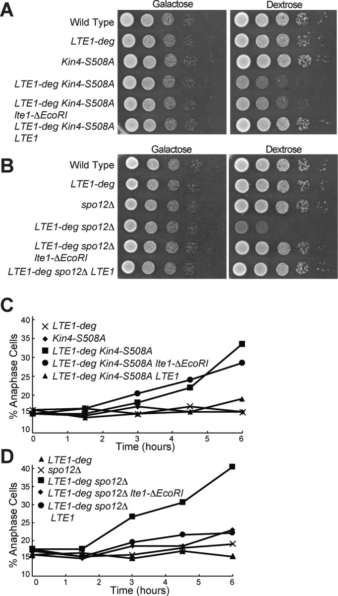
FIGURE 8:

The GEF domains of Lte1 promote exit from mitosis. (A, B) Tenfold serial dilutions of overnight cultures of wild type (A2587), pGAL1-10-URL-3HA-LTE1 (A23686), KIN4-S508A (A21299), pGAL1-10-URL-3HA-LTE1 KIN4-S508A (A24084), pGAL1-10-URL-3HA-LTE1 KIN4-S508A pLTE1-lte1ΔEcoRI (AS336), pGAL1-10-URL-3HA-LTE1 KIN4-S508A pLTE1-LTE1 (A38247), spo12Δ (A4874), pGAL1-10-URL-3HA-LTE1 spo12Δ (A24543), pGAL1-10-URL-3HA-LTE1 spo12Δ pLTE1-lte1ΔEcoRI (AS338), and pGAL1-10-URL-3HA-LTE1 spo12Δ pLTE1-LTE1 (A38260) cells were spotted onto YEP plates containing either raffinose and galactose or glucose and incubated at 30°C. (C, D) pGAL1-10-URL-3HA-LTE1 (A23686), KIN4-S508A (A21299), pGAL1-10-URL-3HA-LTE1 KIN4-S508A (A24084), pGAL1-10-URL-3HA-LTE1 KIN4-S508A, pLTE1-lte1ΔEcoRI (AS336), pGAL1-10-URL-3HA-LTE1 KIN4-S508A pLTE1-LTE1 (A38247), spo12Δ (A4874), pGAL1-10-URL-3HA-LTE1 spo12Δ (A24543), pGAL1-10-URL-3HA-LTE1 spo12Δ pLTE1-lte1ΔEcoRI (AS338), and pGAL1-10-URL-3HA-LTE1 spo12Δ pLTE1-LTE1 (A38260) cells were grown overnight in YEP medium with raffinose and galactose to log phase and diluted into YEPD medium. Cells were grown for 6 h at 30°C, and samples were taken at the indicated times to determine the percentage of anaphase cells (n = 200). Note that the data for C and D are from the same experiment, and so the pGAL1-10-URL-3HA-LTE1 data from C are also shown in D.
