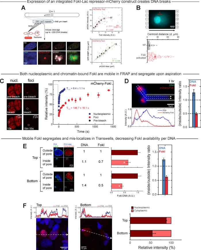
FIGURE 4:
FokI nuclease is confirmed to enact on/off-inducible damage at a specific locus on chromosome 1 and also to segregate/mislocalize during Transwell migration. (A) A lac operator transgene integrated into the p-end of chromosome 1 in the U2OS line. Expression of the integrated mCherry-Lac repressor-FokI construct can be stably induced with 4-OHT (tamoxifen) and Shield-1 to creates DNA breaks. FokI intensity correlates with γH2AX intensity, and the area of GFP-53BP1 at the damaged site is linearly correlated to γH2AX focus area. (B) A comet assay is sensitive enough to detect induced DNA breaks: the mean centroid distance is higher for FokI-activated cells (≥175 nuclei/group; at least three experiments). (C, D) FRAP shows that both nucleoplasmic and chromatin-bound mCherry-FokI are mobile, with half-lives of 6 s and >2 min, respectively. The former is thus much more mobile, and it indeed segregates outside a micropipette in D during aspiration (≥4 cells/group; at least three experiments). (E, F) Migration through 3-μm Transwell pores also causes FokI to segregate and mislocalize, as seen in migrated cells that have been fixed for confocal imaging. Intensity profiles are shown along the dashed arrows (>10 cells/group; at least three experiments).

