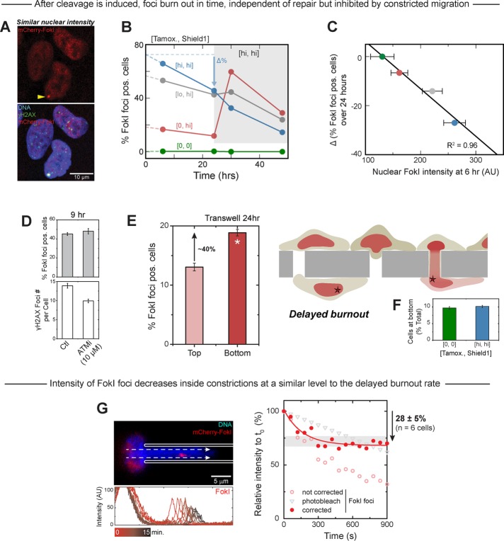
FIGURE 5:
Constricted migration—but not repair inhibition—affects a FokI-cleaved locus on chromosome 1. (A–C) FokI cleavage of the engineered chromosome 1 locus is induced at 0 h by controlled addition of two components (tamoxifen, Shield1). High tamoxifen (1 μM, blue) yields a greater percentage of cells positive for mCherry-FokI foci at 6 h than either low (0.1 μM, gray) or zero (red) tamoxifen. In the absence of both inducing chemicals (green), zero foci develop. For all three high-Shield1 groups (1 μM), the percentage of foci-positive cells decreases between 6 and 24 h even as the nucleoplasmic mCherry signal remains strong, indicating “burnout” of the LacO target site by the nuclease. At 24 h, the FokI construct is reactivated with high tamoxifen in all three high-Shield1 groups. Whereas the high-tamoxifen group continues to burn out as before, there are spikes in FokI foci–positive cells for the low- and zero-tamoxifen groups. The burnout rate between 6 and 24 h is used to extrapolate the percentage of FokI foci–positive cells at 0 h, which is, in turn, used to calculate the change in the percentage of foci-positive cells over 24 h. This ∆% defined in B increases in magnitude with nuclear FokI intensity. Hence high tamoxifen (blue) leads to high nuclear FokI levels and correspondingly greater burnout. Low (gray) and zero (red) tamoxifen, as well as zero tamoxifen/Shield1 (green), show less nuclear FokI and less burnout. At least three experiments. (D, E) Inhibition of ATM kinase, which phosphorylates H2AX to make γH2AX, decreases, as expected, the small mCherry-negative focus numbers per cell, but it does not decrease FokI focus numbers. Burnout is thus limited by nuclease induction rather than by repair. Constricted migration through 3-μm Transwells leads to ∼40% more cells with FokI foci on the bottom than on top (at least three experiments), which suggests that mobile protein loss during migration impedes targeted FokI activity and so delays burnout. (F) Compared to the nontreated cells, activation of FokI expression by 1 μM tamoxifen and 1 μM Shield1 does not impede migration, as shown by the number of migrated cells at the bottom of the Transwell (≥3 Transwells for each group, at least three experiments). (G) A FokI focus drawn into a 3-μm pipette loses ∼30% (≥6 cells) of its initial intensity within minutes, with suitable correction for constant photobleaching.

