
Figure 11.
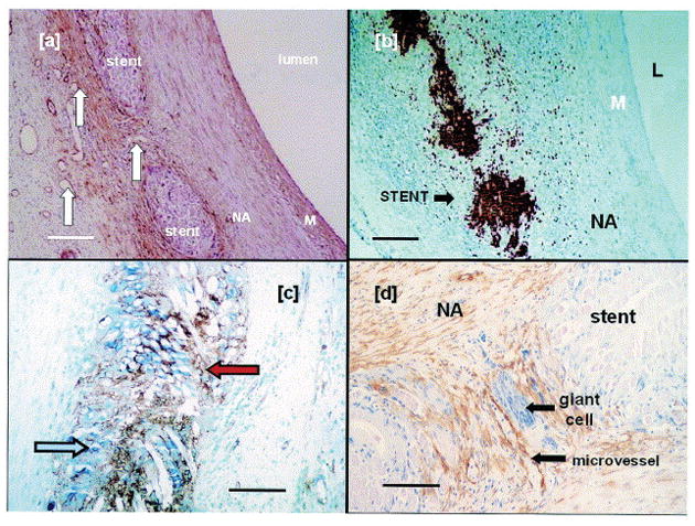
Photomicrographs of pig SVGs fitted with an 8 mm PGA external stent after one month. In and around the PGA sheath, there is a high density of a) microvessels (white arrows), b) macrophages as revealed by immunostaining for MAC387, c) endothelial cells (blue arrow) and proliferating cells (PCNA positive, red arrow). Immunostaining for d) α-actin (brown) and giant cells (purple) reveals that VSMCs and giant cells have also accumulated in the interstitial space. In contrast, microvessels, macrophages, endothelial cells, and proliferating cells are largely absent in the media of the graft. Scale bar for 100 μm for a) and b), and 50 μm for c) and d). Reproduced with permission.[91] Copyright 2004, Elsevier.