
Figure 5.
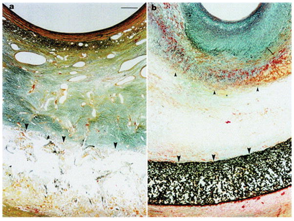
Trichrome staining of porcine SVGs after one month wrapped with a) an 8 mm loos-fitting macroporous Dacron stent and b) an 8 mm loose-fitting microporous PTFE. The internal elastic lamina marked by small arrows and the external elastic lamina marked by large arrows clearly shows a large reduction in NH with the macroporous Dacron stents compared to the PTFE stent. The neointima in the PTFE-wrapped SVGs is highly collagenous, as indicated by the green-blue staining. Orange/brown staining represents VSMCs. There are large, abundant microvessels present in the Dacron-treated grafts, whereas the microvessels in b) are clearly absent, with no large microvessels outside the stent. Scale bare in a) applies to b) as well, and represents 50 mm. Reproduced with permission.[130] Copyright 2001, Elsevier.