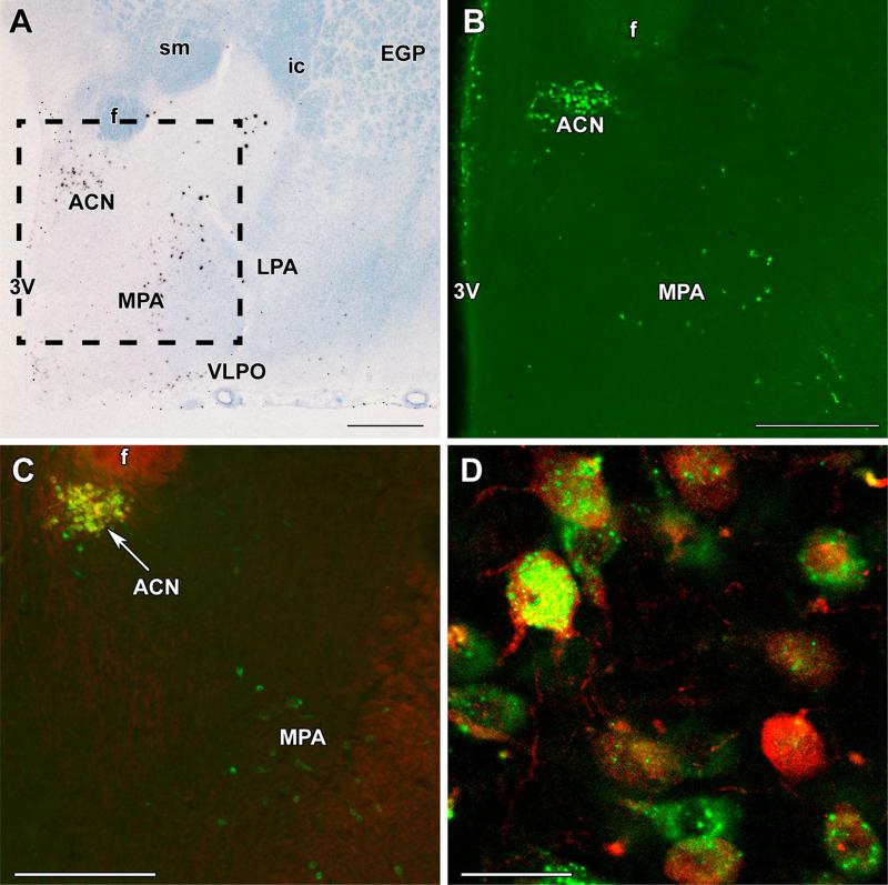
Fig. 1.
Galanin neurons in the preoptic area and their oxytocin content. a A bright-field in situ hybridization histochemistry image at bregma level −0.9 mm demonstrates the presence and distribution of galanin mRNA-expressing cells (black signal) in the preoptic area of a mother rat. b A picture of a galanin-immunolabeled section corresponding to the framed area in a is shown. c Oxytocin immunoreactivity (red) is present in galanin neurons (green) of the anterior commissural nucleus (ACN), but not in the medial preoptic area (MPA). d A high-magnification confocal image demonstrates the double labeling of neurons in the ACN. Note the different distributions of galanin and oxytocin immunoreactivity in the cell bodies and the oxytocin labeling of proximal dendrites. f fornix, ic internal capsule, EGP external globus pallidus, LPA lateral preoptic area, sm stria medullaris, VLPO ventrolateral preoptic nucleus, 3V third ventricle. Scale bars 500 μm for a, 400 μm for b and c, and 40 μm for d

