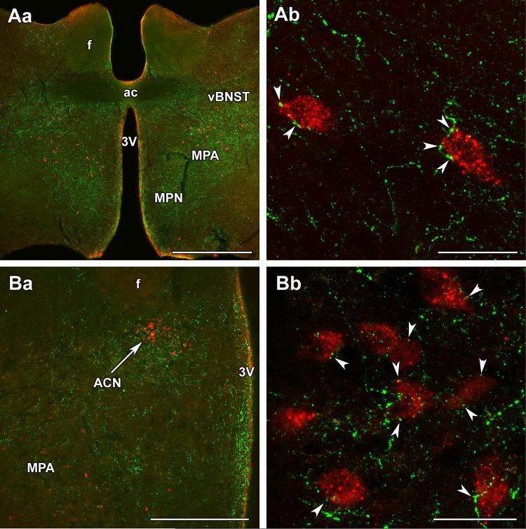
Fig. 3.
Topographical relationship between TIP39 fibers and galanin neurons in the preoptic area of mother rats. Aa TIP39 fibers (green) are located in the medial preoptic nucleus (MPN), medial preoptic area (MPA), and the ventral subdivision of the bed nucleus of the stria terminalis (vBNST) at the caudal level of the anterior commissure (ac). In the MPA, TIP39 fibers are located around galanin neurons (red). Ab A high-magnification confocal image demonstrates that galanin-ir neurons are closely apposed by TIP39 varicosities in the MPA. Ba The anterior commissural nucleus (ACN) is visible in the dorsal part of a more caudal level of the preoptic area. Bb Galanin neurons in the ACN are also closely apposed by TIP39 varicosities (white arrowheads). f fornix, 3V third ventricle. Scale bars 1 mm for Aa, 50 μm for Ab, 500 μm for Ba, and 50 μm for Bb

