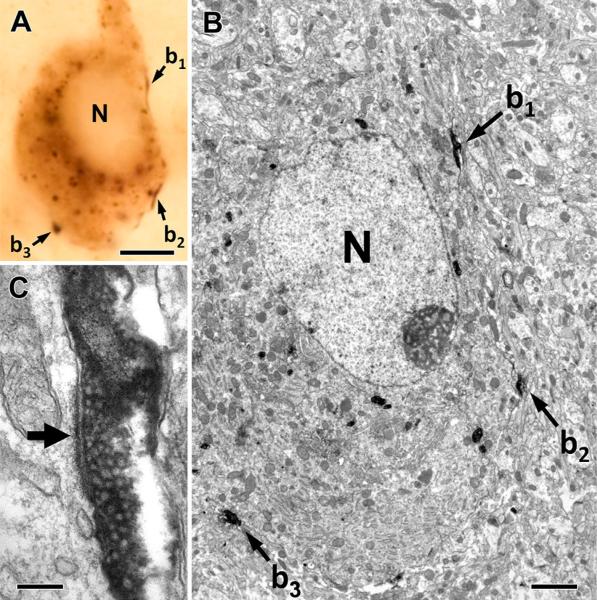
Fig. 4.
TIP39-positive thalamic axons innervate galanin neurons in the MPA. Correlated light and electron micrographs of a galaninimmunoreactive cell that receives three contacts from TIP39-positive thalamic boutons (b1–b3) in the plane of the section. The low-power (a) and high-power micrographs (b, c) verify that the conventional asymmetrical synapses are established on the soma of the galaninimmunoreactive cell as demonstrated by the long post-synaptic specialization (filled arrow) of the synapse formed by a TIP39-positive terminal (b1) in c. N nucleus. Scale bars 5 μm for a, 2 μm for b, and 200 nm for c

