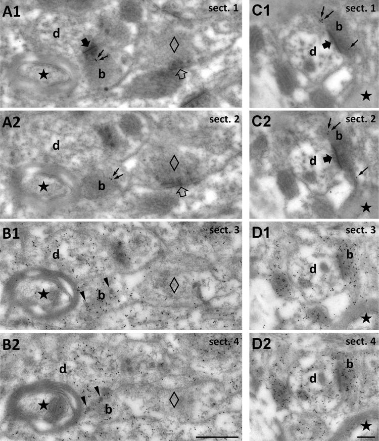
Fig. 5.
Demonstration of the glutamate content of TIP39 terminals in the MPA using consecutive serial electron micrographs sections. a1, a2 Consecutive sections (sect. 1–4) labeled for TIP39 to demonstrate TIP39 immunoreactivity (10 nm gold particles, small black arrows) in a bouton (b) establishing asymmetric synapse (large filled arrow visible only in a1) with a thin, unlabeled dendrite (d). Note that TIP39 immunoreactivity is absent from putative inhibitory terminals (empty diamond) establishing a symmetric synapse (empty arrow) on a dendrite. b Immunogold labeling for glutamate (10 nm gold particles) demonstrates that glutamate is enriched in the TIP39-immunopositive terminal seen in a. The black star shows a myelinated axon as one of the markers that demonstrate that sections in b are consecutive to those in a. In addition, note the presence of dense core vesicles (black arrowheads) implying neuropeptide content in the TIP39-positive terminal. The bouton that establishes symmetric (putative inhibitory) synapse seen in a (empty diamond in all panels) and the myelinated axon (black star) do not accumulate glutamate. c, d Another example of a glutamatergic TIP39 terminal shown in consecutive serial sections. c1 and c2 are immunolabeled for TIP39, while d1 and d2 are labeled for glutamate. Scale bar 500 nm for a and b, and 200 nm for c and d

