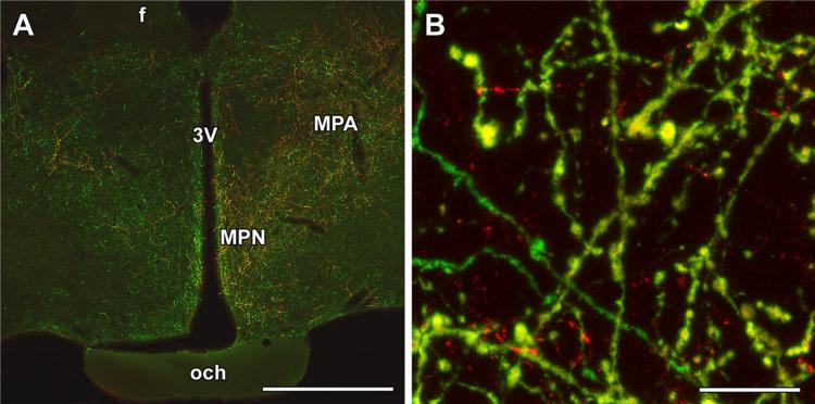
Fig. 7.
Anterogradely labeled fibers in the preoptic area following injection of biotinylated dextran amine (BDA) into the posterior intralaminar complex of the thalamus (PIL). a A high density of BDA-labeled (red) fibers appear in the preoptic area ipsilateral to the injection site, while a few BDA fibers are also present in the contralateral side. The distribution of the anterogradely labeled neuronal fibers is similar to that of TIP39-ir (green) fibers in the preoptic area. b A high-magnification confocal image demonstrates that double-labeled fibers (yellow) are intermixed with the single-labeled ones. f fornix, MPA medial preoptic area, MPN medial preoptic nucleus, och optic chiasm, 3V third ventricle. Scale bars 1 mm for a, and 30 μm for b

